Pregnancy Appointment Schedule: Trimester Guide
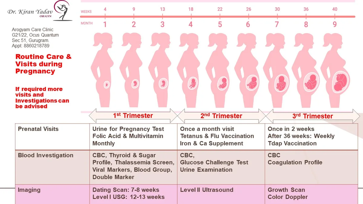
A standard pregnancy appointment schedule by trimester follows a predictable rhythm, but your specific needs might shift those dates. Most providers aim for monthly visits in the first and second halves, then bi-weekly or weekly checks as you approach delivery.
We’ve seen how missing key windows, like the 11 to 14-week nuchal translucency scan, limits diagnostic options permanently. Per ACOG guidelines as of 2026, risk stratification dictates whether you stick to the baseline or need extra monitoring. Let’s break down exactly what happens when, so you don’t miss a critical beat.
Quick Answer
The typical pregnancy appointment schedule by trimester starts with a booking visit at 8-12 weeks. You’ll have monthly checkups until week 28. From week 28 to 36, visits occur every two weeks.
After week 36, appointments become weekly until delivery. High-risk pregnancies require more frequent monitoring. Always confirm your specific timeline with your provider.
Why Your Pregnancy Timeline Isn’t One-Size-Fits-All
Your doctor doesn’t just pick random dates for your checkups. They follow clinical evidence linking specific gestational ages to developmental milestones and potential complications. For example, screening for chromosomal abnormalities works best within narrow timeframes.
Missing these windows can mean losing access to certain non-invasive tests.
Risk factors change everything. If you have pre-existing hypertension, diabetes, or are carrying multiples, your schedule tightens significantly. We often see low-risk patients on a standard monthly track.
High-risk patients might see their provider every two weeks from day one.
Understanding this distinction helps you prepare mentally and logistically. It’s not about being "normal" versus "abnormal." It’s about matching surveillance intensity to biological need. Our research shows that personalized care plans reduce anxiety and improve outcomes by catching issues early.
You’ll also notice variations based on who provides your care. Midwife-led units in the UK often integrate home visits differently than hospital-based obstetric clinics in the US. But the core medical logic remains consistent across ACOG practice bulletins.
The goal is always timely detection and intervention.
The First Trimester: Establishing Baselines and Critical Windows
This phase sets the foundation for your entire pregnancy journey. Your first major appointment, often called the booking visit, usually happens between weeks 8 and 12. Here, we establish your due date using last menstrual period (LMP) data or an early ultrasound measuring crown-rump length.
Blood work is comprehensive during this window. We check your blood group and Rh status to determine if you’ll need RhoGAM later. Infection screenings for HIV, hepatitis B and C, and syphilis happen now.
Early treatment prevents vertical transmission to the baby, reducing risks to near zero.
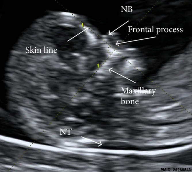
Genetic screening decisions peak here. The nuchal translucency (NT) scan must occur between 11 weeks and 13 weeks plus 6 days. This ultrasound measures fluid behind the fetal neck.
Combined with blood markers, it assesses risk for Down syndrome and other trisomies.
If you opt for cell-free DNA testing (NIPT), it can start as early as 10 weeks. However, NIPT is a screen, not a diagnostic test. Positive results require confirmation via chorionic villus sampling (CVS) or amniocentesis.
These invasive procedures carry small miscarriage risks, so counseling is vital.
Don’t forget physical exams. Your provider will check your weight, blood pressure, and pelvic health. Urine dipsticks test for protein and sugar.
Elevated levels might hint at underlying kidney issues or early gestational diabetes, prompting closer watch in later months.
The Second Trimester: Structural Checks and Glucose Screening
Weeks 14 through 27 are often called the "honeymoon phase" because nausea typically subsides. But medically, this period is heavy on diagnostics. The most significant event is the anatomy scan, scheduled between 18 and 22 weeks.
This detailed ultrasound examines fetal organs, limbs, and brain structure. We also locate the placenta. If it covers the cervix (placenta previa), your delivery plan changes drastically.
C-section becomes likely, and pelvic rest may be prescribed.
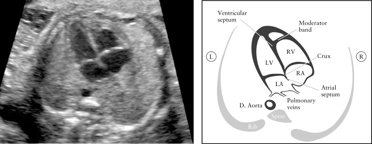
Around weeks 24 to 28, glucose screening begins. Most providers use a one-hour 50g glucose challenge test. If your blood sugar reads high, you’ll follow up with a three-hour oral glucose tolerance test (GTT).
Diagnosing gestational diabetes mellitus (GDM) early allows for diet management or medication, preventing macrosomia (large baby) and birth trauma.
Rh-negative mothers receive RhoGAM immunoglobulin at 28 weeks. This shot prevents your immune system from creating antibodies against Rh-positive fetal blood cells. Without it, subsequent pregnancies could suffer hemolytic disease.
It’s a simple step with profound long-term benefits.
Routine visits continue monthly. We monitor fundal height starting around week 20. By measuring the distance from your pubic bone to the top of your uterus, we estimate fetal growth.
Discrepancies larger than 2-3 centimeters trigger further investigation, such as a growth ultrasound.
The Third Trimester: Growth Monitoring and Delivery Prep
From week 28 onward, the pace accelerates. Visits shift to every two weeks until week 36. Then, they become weekly until delivery.
This frequency ensures we catch rapid changes in blood pressure or fetal movement patterns.
Group B Streptococcus (GBS) screening occurs between 35 and 37 weeks. This bacterial swab determines if you carry GBS. If positive, you’ll receive IV antibiotics during labor.
This single intervention reduces newborn sepsis rates dramatically. It’s painless but critical.
Vaccinations align with this window too. The Tdap vaccine protects against pertussis (whooping cough). Administered between 27 and 36 weeks, it transfers maternal antibodies to the fetus.
These antibodies protect your baby during their vulnerable first few months of life before they can get vaccinated themselves.

Fetal position matters now. At 36-37 weeks, we check if the baby is head-down (cephalic). If breech, we discuss external cephalic version (ECV).
This manual maneuver attempts to turn the baby externally. Success rates hover around 50%, avoiding unnecessary C-sections.
Cardiotocography (CTG) or non-stress tests (NST) may begin if you’re overdue or high-risk. These monitors track heart rate variability alongside uterine activity. Reduced variability or decelerations signal distress, prompting immediate delivery evaluation.
Trust your instincts here; decreased movement warrants an urgent call, not waiting for your next scheduled slot.
How Risk Factors Change Your Appointment Schedule
Not everyone fits the standard mold. Your provider adjusts frequencies based on individual health profiles. Understanding these triggers helps you anticipate more intensive care without feeling alarmed.
It’s proactive medicine, not punitive oversight.
High-risk conditions demand tighter loops. Chronic hypertension requires weekly blood pressure checks and urine protein analysis from early on. Preeclampsia develops quickly, so vigilance saves lives.
Similarly, pre-gestational diabetes necessitates frequent glucose monitoring and fetal echocardiograms to check heart development.
Multiple gestations (twins or triplets) automatically bump you into higher surveillance. Twin pregnancies often involve separate placentas or shared ones. Monochorionic twins face unique risks like twin-to-twin transfusion syndrome.
Ultrasounds may happen every two to four weeks throughout the second and third trimesters.
Advanced maternal age (over 35) increases chromosomal anomaly risks. While NIPT is available, some providers offer earlier CVS or additional growth scans. BMI over 30 complicates ultrasound visibility and raises GDM risks.
Expect more frequent glucose testing and potentially earlier induction discussions.
Previous adverse outcomes matter too. A history of stillbirth, preterm birth, or recurrent miscarriage leads to customized protocols. Cervical length measurements via transvaginal ultrasound might start at 16 weeks.
Progesterone supplementation or cerclage placement could be considered to prevent preterm labor.
| Risk Factor | Standard Visit Frequency | Adjusted Frequency | Key Additional Tests |
|---|---|---|---|
| Low-Risk Singleton | Monthly (wks 8-28) Bi-weekly (wks 28-36) Weekly (wks 36+) | None | Routine labs, Anatomy scan |
| Gestational Diabetes | As above | Weekly/Bi-weekly from wks 24 | Serial growth scans, NSTs |
| Chronic Hypertension | As above | Weekly from wks 16 | Protein/Creatinine ratio, Dopplers |
| Twin Pregnancy | As above | Every 2-4 weeks wks 16-36 | Cervical length, Detailed anatomy |
| Previous Preterm Birth | As above | Bi-weekly cervical checks wks 16-24 | Vaginal progesterone consideration |
These adjustments aren’t arbitrary. They stem from data showing improved neonatal survival rates with intensified monitoring. If your provider suggests extra visits, view them as safety nets rather than signs of trouble.
Communication is key. Ask why each addition is necessary. Ensure you understand the specific marker being watched.
This partnership empowers you to advocate for appropriate care while staying calm amidst increased medical attention.
Common Scheduling Mistakes That Miss Vital Screens
The biggest error isn’t skipping a visit. It’s booking the wrong test at the wrong time. The nuchal translucency window closes strictly at 13 weeks and 6 days.
If you wait until week 14, that specific screening option disappears. You might rely solely on NIPT or later anatomical scans, losing valuable early data.
Another frequent slip is delaying the glucose challenge test. Many patients assume they can wait until symptoms appear. Gestational diabetes often presents silently.
Missing the 24 to 28-week window means undetected high blood sugar affects fetal growth. This leads to macrosomia and complicated deliveries.
Procrastinating on Rh immunoglobulin is risky too. The shot must be given by 28 weeks for Rh-negative mothers. Waiting until labor starts leaves no time for antibody prevention.
Once sensitization occurs, it’s permanent. Future pregnancies face severe hemolytic disease risks.
Don’t ignore missed appointments due to logistics. Life gets busy, but prenatal slots are finite. Rescheduling a critical anatomy scan pushes it past optimal visualization limits.
Fetal position changes, making details harder to see. Always prioritize these dates over convenience. Communicate early if conflicts arise.
Providers can often squeeze you in before the deadline passes.
When to Call Your Provider Between Appointments
Routine checkups don’t cover emergencies. You need clear triggers for unscheduled contact. Decreased fetal movement is the top warning sign after 28 weeks.
If you feel fewer than ten kicks in two hours, call immediately. Don’t wait for your next weekly visit. Time matters in potential distress scenarios.
Vaginal bleeding requires prompt evaluation. Spotting can be normal, especially after intercourse. However, bright red flow or clots signal placental issues.
Previa or abruption demands urgent assessment. Even painless bleeding warrants a same-day call. Better safe than sorry with placental integrity.
Severe headaches paired with vision changes indicate preeclampsia. Standard morning sickness nausea differs from sudden onset vomiting. Watch for upper right abdominal pain too.
These symptoms suggest liver involvement. Blood pressure spikes above 140/90 mmHg need reporting. Home cuffs help, but clinical verification is essential.
Fluid leakage is another red flag. Distinguishing urine from amniotic fluid can be tricky. Use pH test strips if provided.
Persistent wetness suggests ruptured membranes. Infection risk rises quickly after rupture. Early antibiotics or delivery planning prevents complications.
Trust your instincts. If something feels off, speak up. Your provider expects these calls.
They’d rather evaluate false alarms than miss true emergencies. Prompt communication protects both you and the baby.
Frequently Asked Questions About Prenatal Visits
How many prenatal visits are typical?
Most low-risk pregnancies involve 10 to 14 visits. This includes the initial booking appointment. Subsequent checks happen monthly until week 28.
Then bi-weekly until week 36. Finally, weekly visits continue until birth. High-risk cases may exceed 20 appointments.
Frequency depends on maternal health and fetal development progress.
Can I skip my first trimester ultrasound?
You shouldn’t skip it without medical advice. The dating scan confirms viability and corrects gestational age errors. It identifies ectopic pregnancies or multiple fetuses early.
Missing this step delays critical care decisions. Insurance often covers it as preventive care. Accurate dating ensures all future screenings align correctly.
Skipping creates diagnostic blind spots.
What happens if I miss a glucose test?
Contact your provider immediately. Late testing reduces accuracy for gestational diabetes diagnosis. You may need an oral glucose tolerance test instead.
Unmanaged blood sugar harms fetal growth. Catching GDM late limits dietary intervention windows. Follow-up monitoring becomes more intensive.
Never ignore this screening window. It directly impacts delivery safety and newborn health outcomes.
Are home births included in standard schedules?
Home birth protocols differ significantly. Midwives typically visit less frequently in hospitals. Instead, they conduct more extensive home assessments.
Emergency transfer plans are crucial. Not all providers support home births. Check local regulations and insurance coverage.
Hospital-based care offers immediate access to surgical teams. Ensure your chosen model matches your risk profile perfectly.

Similar Posts